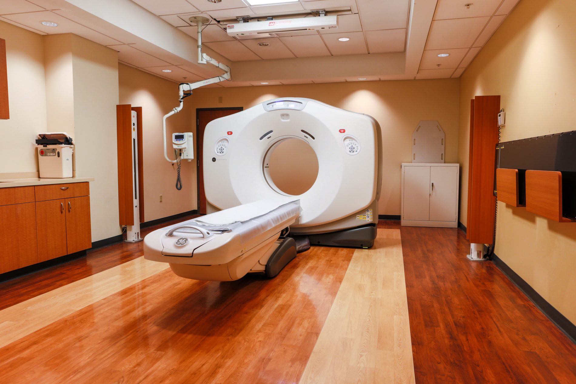
Medical imaging procedures, such as computed tomography scans (CT) and positron emission tomography scans (PET), are used to identify and diagnose many different cancers and conditions. Zangmeister Cancer Center continues to invest in next-generation technologies that provide precise views of internal organs, bones, soft tissues and blood vessels with immense clarity and detail. These scans and other imaging tests are performed and reviewed on-site.
Frequently Asked Questions
The following information will help you understand and prepare for your exam. Our staff and your physician will also be able to answer additional questions you may have.
What is a CT scan?
A Computed Tomography (CT) scan uses a combination of x-rays and computers to give the radiologist an advanced but non-invasive way to view your body’s internal anatomy. A CT scan rapidly acquires 2-dimensional pictures of your anatomy which are then reconstructed by a computer into 3-dimensional images for in-depth clinical evaluation.
What is a PET/CT scan?
A PET/CT combines functional information from a positron emission tomography (PET) exam with anatomical information from a CT scan into one single exam. A PET scan detects changes in how your cells are using nutrients such as sugar and oxygen. Since these functional changes often take place before physical changes occur, PET can provide information that often allows your physician to make an earlier diagnosis.
The PET exam identifies metabolic activity in the cells, and the CT scan provides an anatomical reference or “roadmap”. When these two scans are fused together, physicians can view metabolic changes seen on PET in the proper anatomical context with your body as seen on the CT scan.
Why do I need this exam?
Your exam results may have a major impact on your physician’s diagnosis of a potential health problem and how a treatment plan is developed and managed in the event that a health issue is discovered. These tests not only help your physician diagnose a problem, they also help establish the extent and severity of that disease. Additionally, scan results help predict the likely outcome of various treatment alternatives, identify the best approach for treatment and help monitor your progress. That way, if you’re not responding as well as expected, you can be switched to a more effective treatment as soon as possible.
How should I prepare for my exam?
Please follow the instructions below to ensure the most accurate results.
CT (CAT scan) Preparation
Abdomen/Pelvis:
- Nothing to eat or drink except water after midnight or 4 hours prior to exam.
- Please drink as much water as you can the day of your scan just prior to your appointment.
All Other Contrast CT’s:
- Nothing to eat or drink except water 4 hours prior to exam.
PET/CT Preparation
- Refrain from heavy exercise 48 hours prior to exam.
- Eat a low‐carb meal (no sugar, fruit, breads, desserts) for the 2 meals before your exam and drink extra water the night before and the day of the exam.
- You are required to fast 4 hours prior to your exam (no food, drinks, snacks, candy, mints, etc., other than plenty of water).
- You may take any medication you need to take with water.
- If you are a diabetic, please notify us upon arrival at the imaging center.
- Wear warm comfortable clothing preferably with no metal (zippers, rivets) or jewelry.
- Arrive on time for your appointment to ensure the injectable glucose does not expire.
What should I expect when I arrive?
We will review your history and past exams. Depending on your procedure you may be asked to drink an oral contrast to help visualize your digestive tract. If you are having a PET/CT scan we will also take a small blood sample to ensure your blood glucose levels are within normal limits. If a CT scan was requested by your physician, we will also determine if your kidneys are functioning normally by measuring certain chemicals in your blood sample.
For the PET scan, you will receive an injection of a radioactive tracer similar to glucose in the vein. This material must pass multiple quality control measures before it is used for any patient injection. In all cases, the radioactivity diminishes rapidly from the body such that most of it is gone from your body within 12 hours of injection and ALL of it is gone within 24 hours.
For most PET Scan studies you will wait one hour for the glucose-like injection to distribute itself evenly throughout your body. During this time, you will be asked to relax in an easy chair. If your physician has requested that you have a contrast CT then we will also place a small IV catheter in a vein. This will allow the technologist to administer the IV contrast during your CT scan. The IV catheter is immediately removed following the procedure.
What will the scan be like?
You will lie on a comfortable but narrow table on a scanner. The table will move slowly through the short tube-shaped scanner as it acquires the images. You will be asked to lie very still during the scan because movement can interfere with the results. During your scan you might hear a slight humming sound, but you will not feel anything unusual. You may feel the table move as it acquires images it needs from various positions.
If a CT scan is performed with IV contrast, you may feel a slight warmth throughout your body as well as a metallic taste. This is normal and should not alarm you. The technologist will monitor you during the exam. More specific details of your scan will be explained fully by the technologist upon your arrival.
How long will all this take?
A typical CT scan can take from 10-20 minutes, depending on the procedure performed. However, if oral contrast is administered, you will be asked to relax and wait an additional 30 minutes for the contrast to fully distribute though your digestive system. The exam procedure can vary depending on what we are looking for and what we discover along the way.
The PET/CT scan itself should last between 15-20 minutes, however, plan to spend at least 2 hours as this will include the time prior to the scan necessary for the radiotracer to be distributed throughout your body.
What happens after my exam?
You may leave us as soon as your exam is complete. Unless you have received special instructions, you will be able to eat and drink immediately – drinking lots of fluids after your exam will help remove any of the radioactive glucose and/or contrast material that may still be in your system. In the meantime, we’ll begin preparing results for review by our radiologist who will then pass along these results to your physician. Your physician will then explain the results of the test to you.
Safety of PET/CT exams
Be assured that both the PET/CT and CT exams are safe and effective diagnostic procedures. Although both involve exposure to diagnostic level radiation, your physician has determined that the risk of this exposure outweighs the risk of not having the scans and thus the information the scans can provide. The radiopharmaceuticals used in a PET/CT scan do not remain in your system for long so there is no reason to avoid interacting with other people once you have left our facility. To be extra safe, however, wait 12 hours before getting too close to an infant or to someone who is pregnant.
Rarely, patients may experience an allergic reaction to the IV contrast used for CT scans. If you have experienced any previous reactions to IV contrast, iodine, or seafood please notify our center prior to your appointment so we can discuss alternative preparation options with you and your physician.
Restrictions
Please let our staff know prior to your test if you are pregnant or think you might be pregnant. While these radiation levels are acceptable for you, they may not be for a developing fetus. If you are diabetic, please monitor your blood sugar closely prior to your exam. If your blood sugar levels are above 180 on the day of your exam, please contact our facility as soon as possible. If your blood sugar is too high, we may not be able to perform your PET/CT exam.


